Liver cells divvy up metabolic tasks to take advantage of available nutrients.
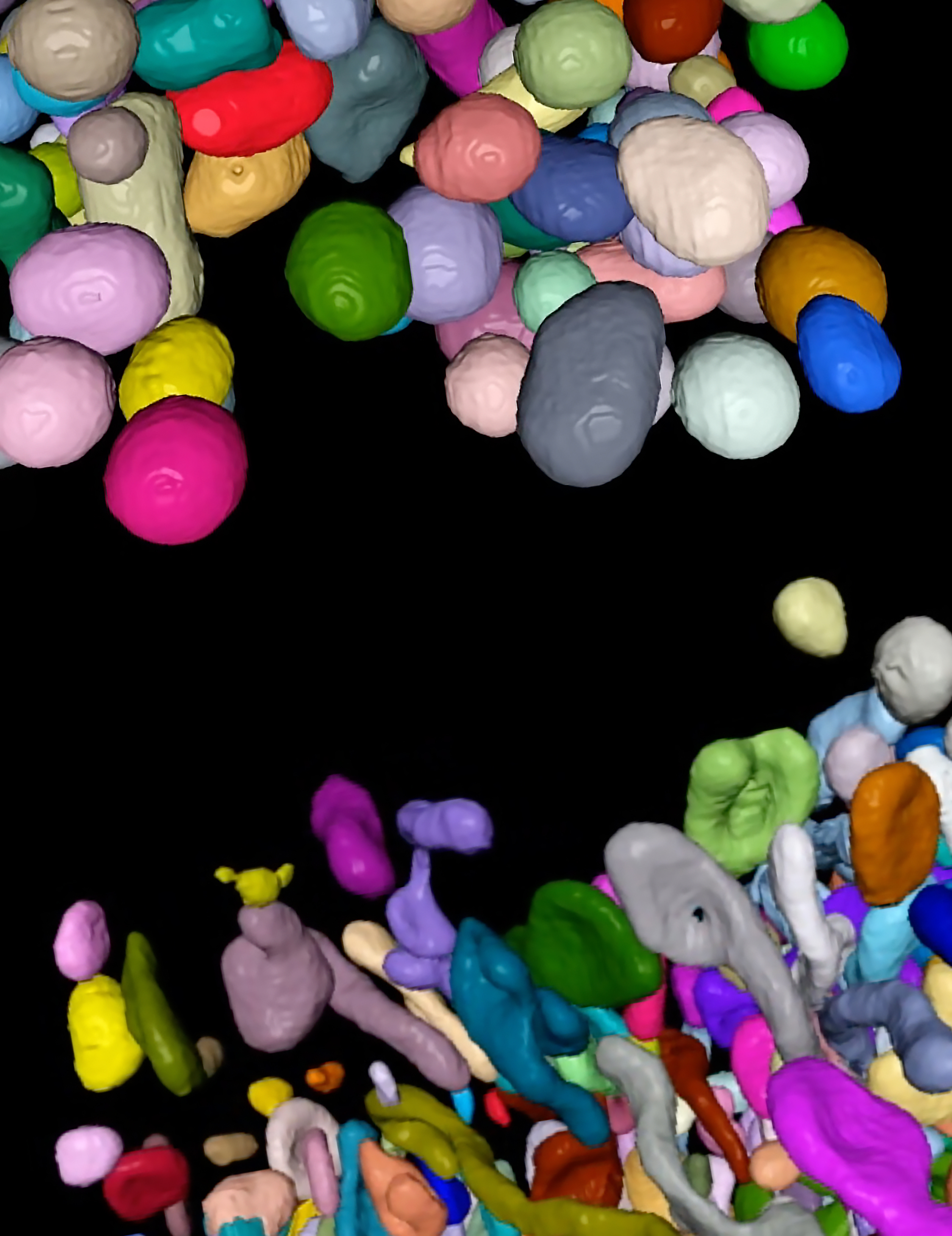
At the top of this image is a depiction of mitochondria from liver cells at the edges of liver lobules. These mitochondria typically have access to nutrient-rich blood and convert some of these nutrients to energy. At the bottom is a depiction of mitochondria from liver cells closer to the center of liver lobules. These mitochondria focus on equipping cells to synthesize lipid molecules for later use. CCR researchers have uncovered new details on the relationship between mitochondria function, structure and position in the liver, with implications for human health and disease.
Credit: Adam Harned and Kedar Narayan, CCR, NCI, NIH; SPGM, FNL, NCI, NIH
The liver is a multitasking organ, responsible for clearing toxins from the blood, breaking down nutrients, generating energy, storing fats and hundreds of other functions. Every cell has a job to do and is equipped with specialized tools to do it. Stadtman Tenure-Track Investigator Natalie Porat-Shliom, Ph.D., is examining how and why liver cells adopt their particular features and functions. She wants to understand this cellular diversity in healthy livers so she and others can make sense of changes that occur during disease.
Porat-Shliom explains that a quarter of adults in the developed world are affected by a progressive spectrum of diseases related to fat buildup in the liver. As the prevalence of these diseases increases, they are becoming a leading cause of liver cancer. One hallmark of this type of liver disease is changes to structures inside liver cells called mitochondria. To understand the significance of these changes, Porat-Shliom and her team think it is important to first fully appreciate natural variations in mitochondria’s form and function inside the liver.
The liver is organized into thousands of working units known as lobules. Some of the cells that make up each lobule have mitochondria that appear large and round under a microscope, whereas others are elongated like noodles. In the livers of healthy, well-fed mice, the round mitochondria appear at the edges of each lobule, where nutrient-rich blood enters. In the center of the lobules, where blood exits to a central vein, mitochondria adopt their skinnier form.
In a study reported in Nature Communications, Porat-Shliom and her team analyzed mitochondria isolated from these different locations in the liver and uncovered new details on the relationship between mitochondria function, structure and position.
They found that the round and skinny mitochondria housed different sets of proteins, suggesting that they perform different functions and hinting at the mechanisms they use to become specialized.
With a series of experiments assessing energy production and storage, the researchers confirmed that at the edges of a liver lobule, where blood arrives rich with nutrients, cells use their mitochondria to convert some of those nutrients to energy. Nearer to the center of the lobule, mitochondria focus instead on equipping cells to synthesize fatty molecules called lipids that can be stored for later use.
The researchers also found that if they blocked liver cells’ ability to sense how many nutrients were available, they could shift the function of the mitochondria in those cells. That suggests liver cells can adjust their metabolic activities to manage critical resources.

“When you have enough nutrients, your liver can multitask,” Porat-Shliom says. “You can afford to make lipids and send them for storage for times when food becomes unavailable.”
Porat-Shliom says her team has seen the same spatial variations in mitochondrial architecture within samples of healthy human liver tissue, and understanding this diversity is critical for shedding light on metabolic diseases that place the liver at a greater risk for cancer. “The liver is just a remarkable organ, and we have the technology now to really look at this in an unprecedented resolution,” she says.