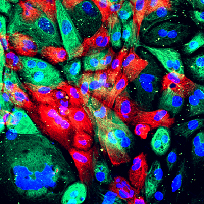
A new imaging method enables sensitive detection and monitoring of high-risk prostate cancers.

Wild-type human prostate cells from an organoid (a man-made construct that resembles an organ). These cells have come from a xenograft where they serve as controls for the study of primary prostate cancer tumor cells, which are also injected into mice and then extracted for characterization. Credit: NCI, NIH
Early detection and treatment of prostate cancer has the potential to save lives. On the other hand, many prostate tumors never become life-threatening. For men with slow-growing prostate cancers, aggressive treatments and their serious side effects may be unnecessary. Thus, the major challenge in prostate cancer diagnosis is not in finding every tumor but in identifying those that are most likely to cause harm if left untreated.
The standard approach to biopsying prostate cancer is to use an ultrasound probe to visualize the prostate. Ultrasound images enable the physician to see where the biopsy needle enters the tissue and to safely and systematically collect tissue samples from throughout the prostate. However, ultrasound does not give a sufficiently detailed picture of the prostate for physicians to biopsy specific lesions, even when they have been detected separately by magnetic resonance imaging (MRI). Thus there is significant potential to miss clinically important tumors.
Advanced imaging technology is changing clinical practice and allowing physicians to perform much more precise prostate biopsies. The CCR’s Urologic Oncology Branch Investigator, Peter Pinto, M.D., along with the Program Director of the Molecular Imaging Program, Peter Choyke, M.D., and the Director of the National Institutes of Health’s Center for Interventional Oncology, Bradford Wood, M.D., are at the forefront of developing high-resolution imaging methods for prostate biopsy. Together they have pioneered the use of ultrasound combined with MRI for biopsies from the laboratory to the clinic. Images collected with the two techniques are super-imposed on one another during the biopsy, which allows a urologist to directly guide the biopsy needle to areas of the prostate that appear suspicious.
In a 2015 study, the team determined that MRI-ultrasound fusion-guided procedures find more high-risk tumors, those that are most likely to grow quickly and spread, than standard biopsies. In 2016, new studies provided further evidence that fusion-guided biopsy can improve prostate cancer diagnosis.
In April, the team reported in the Journal of the National Cancer Institute that combining MRI and ultrasound images makes prostate cancer biopsies more efficient and requires fewer tissue samples than a standard biopsy to detect high-risk cancers. They analyzed biopsies from more than 1,000 men who received both standard and targeted biopsies as part of a clinical trial. Both biopsy methods detected a similar number of cancer cases in the group, but targeted fusion biopsies detected more high-grade cancers than the standard procedure.
Additionally, whereas standard biopsies usually involved the removal of 12 tissue samples, the targeted procedures took an average of only five samples. This is significant because even though prostate biopsies are generally considered safe, they can cause bleeding or infection, and decreasing the number of tissue samples needed to diagnose cancer can reduce the occurrence of these complications.
After further analysis of their clinical trial data, Pinto, Wood, Choyke and their colleagues determined that fusion imaging improves clinicians’ ability to monitor existing prostate tumors and determine whether they have progressed. In a study reported in the Journal of Urology, the team analyzed data from 166 patients with low- or intermediate-risk prostate tumors that were under “active surveillance.” Fusion-guided biopsies detected 26 percent more cancer progression than standard biopsies.
This groundbreaking technology is becoming increasingly available at cancer centers in the United States. The new studies suggest that, because of its ability to distinguish prostate tumors that need treatment from those that do not, it will improve the quality of life for many patients and has the potential to become the standard of care.
Siddiqui MM, et al. J Natl Cancer Inst. 2016 Apr 29;108(9).
Frye TP, et al. J Urol. 2016 Sep 6. doi:pii: S0022-5347(16)31209-5.


