The Laboratory of Cancer Biology and Genetics (LCBG) manages and supports several shared resources to provide our investigators with access to cutting-edge technology and specialized expertise. These in-house resources foster collaboration and enhance the productivity, breadth, and impact of our research program.
LCBG Microscopy Facility
Manager: Ross Lake
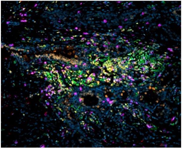

The Laboratory of Cancer Biology and Genetics (LCBG) Microscopy Facility provides training, guidance, and state-of-the-art service to LCBG scientists as well as other NCI-CCR scientists who require whole slide scanning, widefield, or confocal microscopy imaging as part of their research efforts.
Please visit the LCBG Microscopy Facility website to learn more.
LCBG Animal Model and Genotyping Facility
Manager: Wendy Dubois
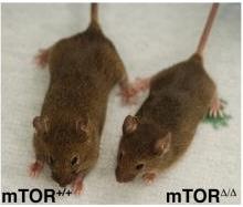

The LCBG Animal Model and Genotyping Facility provides outstanding, reliable research support to LCBG and other NCI intramural investigators. The Facility's mission is to create and maintain mouse models for various cancers including: B cell lymphomas, plasma cell tumors, myeloma, skin carcinogenesis and melanoma, breast cancer, and prostate cancer. The Facility also provides animal study technical support (weaning, genotyping, injection administration, tumor measurement, tissue collection, and surgery) and assistance with animal study proposal submission.
Learn more about the Animal Model and Genotyping Facility.
LCBG High-Dimension Data Analysis Group
Manager: Maxwell Lee, Ph.D.
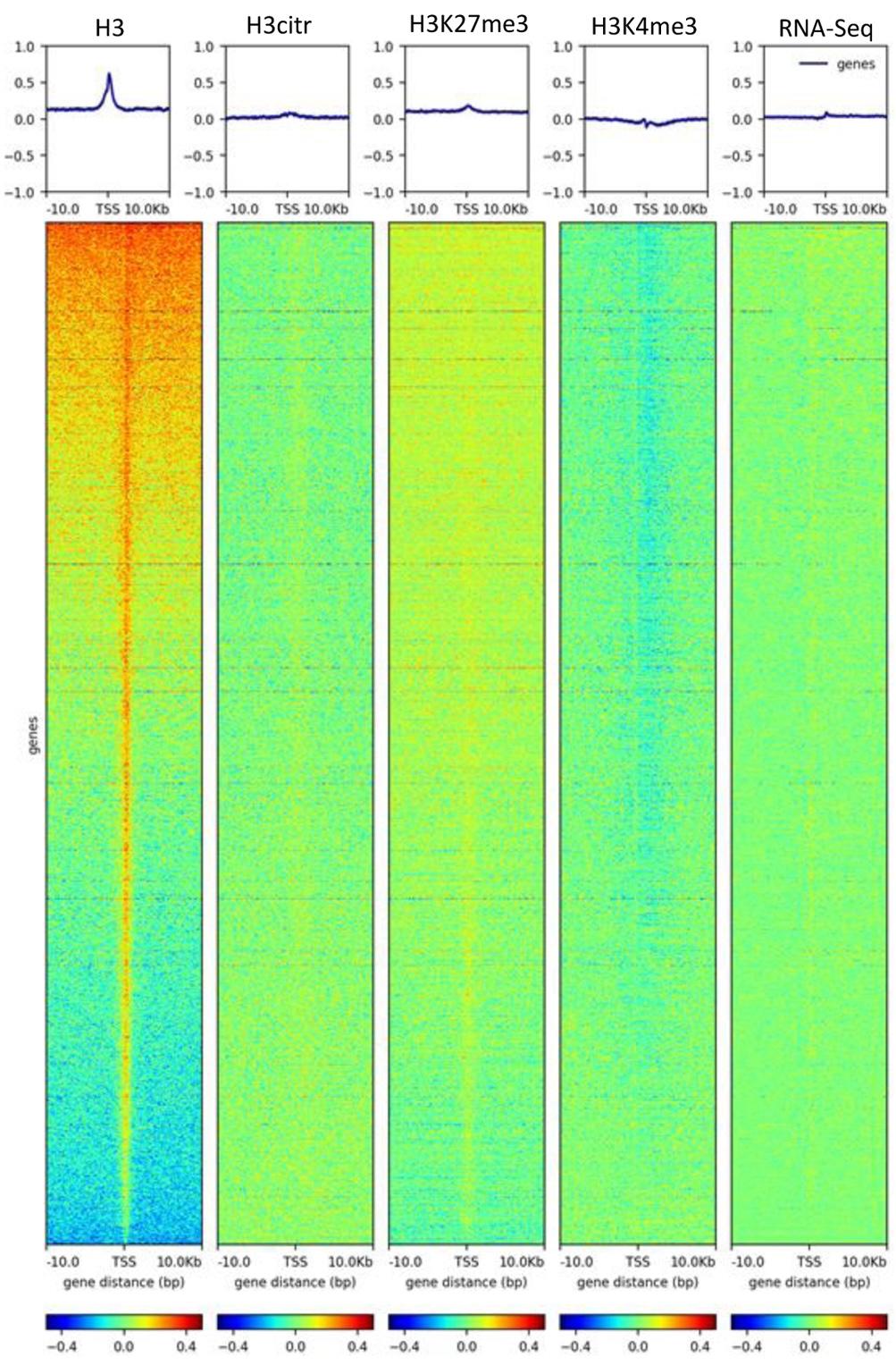

The LCBG High-Dimension Data Analysis Group collaborates with LCBG investigators seeking bioinformatic support, including data analysis, development of analytic tools, result interpretation, study design, and training/education. Data types include whole genome sequencing, exome sequencing, bulk RNA-seq, single-cell RNA-seq, ChIP-seq, ATAC-seq, Methyl-seq, proteomics (RPPA), metabolomic, and microarray. Many types of analysis are supported, including:
- Differential gene expression
- PCA/UMAP/heatmap visualization
- Pathway/network analysis
- Survival analysis, clinical associations
- Somatic and germline mutation analysis
- DNA copy number analysis
- Structural variation analysis
- RNA fusion transcript analysis
- Genome-wide association studies (GWAS) and epidemiology studies
- Phylogenetic and population genetics
- Constructing custom reference genomes
- Genetic regulatory network analysis
- Evolution dynamics
- Epigenomics
- Mathematics and statistical modeling
Learn more about the High-Dimension Data Analysis Group.
LCBG Molecular Pathology Unit
Manager: R. Mark Simpson, D.V.M., Ph.D.
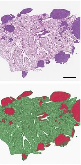

The LCBG Molecular Pathology Unit (MPU) provides opportunities to bridge basic and clinical research efforts by more precisely optimizing the development, characterization, and utilization of models of human disease. The MPU has extensive research and teaching expertise in comparative and investigative pathology and frequently collaborates with LCBG investigators to support:
- Training in comparative and molecular pathology
- Research investigation and animal model validation
- Developing molecular diagnostics and digital pathology for research
- Noninvasive medical imaging
Learn more about the Molecular Pathology Unit.
CCR Confocal Microscropy Core
Manager: Michael J. Kruhlak, Ph.D.
The CCR Microscopy Core supports the microscopy and digital imaging needs of investigators studying the biological structures and cellular processes involved in the cell biology of cancer by applying our diverse imaging resources, expertise, and quality customer service. The goal is to provide cutting-edge imaging technology, expert consultation and training, image analysis, and data management for investigators to advance their cancer research.
Please visit the CCR Confocal Microscopy Core website to learn more.