We offer investigators the latest instrumentation and expertise.
Cryo-EM Facility
in the Center for Structural Biology
Cryo-EM Facility
About
Mission and Scope
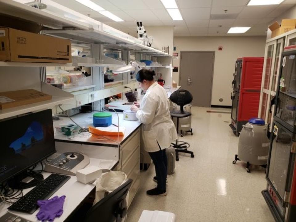
Image
The CSB Cryo-EM Facility began as a Structural Biophysics Laboratory (which later became part of CSB) facility in 2019, with the Talos Arctica fully operational. It was significantly expanded and formally commissioned by Dr. Susan M. Lea in 2022, after she joined NCI from Oxford in 2020. It is equipped with state-of-the-art cryogenic electron microscopes and the most advanced specimen preparation equipment, including a Titan Krios G4 with Selectris/Falcon 4i, a Talos Arctica G2 with Gatan BioQuantum/K3, and an Aquilos II with iFLM and Chameleon. The Titan Krios G4 and Talos Arctica G2 can collect cryo-EM SPA data at up to 900 and 450 movies per hour, respectively. The Aquilos II is used to prepare cryo-ET lamellae samples. A full list of microscopes and specimen preparation devices can be found at Instrumentation.
Our mission is primarily to serve the CSB and CCR community, but microscope time is also available to other NCI and NIH intramural research groups; scientists whose research projects are approved by the review committee can access all equipment in this facility. Non-experts can be trained in sample preparation and cryo-EM operation and collaborations can be established to allow structure determination. For detailed information and procedures on how to access this facility, see User Policies.
Procedure for access
The workflow for access and various forms can be found through the Microscope Access Workflow link.
Training
After the initial proposal is approved, the facility offers training in preparing negative staining samples; operating plunge-freezer equipment (Vitrobot mark 4 and 5, Leica EMGP2, Chameleon; operating standard electron microscope and cryo-EM for screening samples and/or data collection.
Aquilos 2 operation will be trained on-site by a Thermo Fisher Scientific application engineer.
General considerations for access to Arctica and Krios microscopes
A. Talos L120C: Well-trained user(s) can use the microscope to screen negative staining to assess the feasibility of the next step of experiments. These include particle homogeneity, aggregation level, and particle density etc. It should be noted that for many samples, information gained from negative stain does not necessarily translate to success for vitrification and successful grid making.
B. Access to Talos Arctica G2: after confirming that the majority of particles are homogenous and have minimum particle aggregation, users can prepare frozen-hydrated samples to use Talos Arctica G2. This cryo-EM microscope can be used for screening frozen-hydrated grids, small-scale data set collection, and large-scale data collection for structure determination.
C. Access to Titan Krios G4: Access to this microscope is through the following vetting process. First, submit to the committee a brief, updated proposal for approval, including a short progress report demonstrating that the initial results obtained from Arctica meet the following criteria:
- Atlas to show overall ice coverage
- Square image for checking optimized ice thickness
- High-magnification images for particle distribution in holes
- 2D classification or 3D density map with better than 0.4 nm resolution
- Particle distribution plots and 3D-FSC/cFAR scores, a very important metric, should also be provided. A 4 Å anisotropic map on the Arctica will not improve in resolution or quality on the Krios if the particle distributions are limited.
- A reasonable estimation of how much data is needed to solve the structure. The allocated time will be decided by the committee.
- A small data set collection is necessary for identifying specimen quality when 3D density map resolutions could not reach 0.4 nm, but better than 0.7 nm for some difficult samples.
- Users should explore all alternative approaches to improve particle distributions (i.e. adding a surface to the grids, adding detergents to samples, trying the chameleon, etc) before relying on tilted data, as there is a significant decrease in data quality when tilting the stage.
Notably, it is a common practice by the facilities on the Bethesda campus that if a sample giving 4 Å resolution of an electron density map achieved on an Arctica will not be improved to 3 Å and should not be considered for Krios time. Nevertheless, for small particles (< 100 kDa), the Krios can result in better than 1 angstrom map difference across the two microscopes. Furthermore, special consideration may be given to scientific cases where local structural details of components of very large architectural assemblies are known, but the global organization of the components is unknown and is the focus of biological importance; the improved electron density map acquired from Krios may be critical for locating and orienting components within the global architectural structures. In such cases, allocating Krios time can be considered. Moreover, it is noteworthy to mention that there are odd cases where a significant improvement in map quality may be achieved going from the Arctica to the Krios. But all of the metrics above (good 2D classes, good particle density, etc) need to be fulfilled before considering the Krios time.
Access priority and operation policy
The criteria for consideration of access:
A. Feasibility. The feasibility is judged based on the quality of grids screened on Talos L120C (negative staining) and Arctica.
B. Scientific merit and significance.
Ranking of the priority in order given the same scores of the projects:
A. Tenure-track investigators and recently (within 3 years) tenured investigators
B. Senior investigators.
C. The CSB PIs have higher priority over non-CSB PIs, given the same ranking scores of feasibility and scientific merits among different categories of PIs.
Assistance by a cryo-EM specialist
For investigators who do not have the cryo-EM expertise required for conducting rigorous structural biology projects, the CSB cryo-EM facility offers assistance via close interaction with a cryo-EM specialist whose main responsibility is to assist/collaborate with non-cryo-EM PIs with their structural biology projects.
Acknowledgments
Screening cryogenic grids, large-scale data collection, and analyzing negative stain sample results should be acknowledged in publications that use CCF-generated data.
Collaborations (co-authorship)
PIs without cryo-EM expertise could gain access to the microscopes through collaborations with either one of the CSB PIs or the CCF’s dedicated cryo-EM specialist. The following constitutes collaborations: screening negative-stained samples, preparing cryogenic specimens and optimizing cryogenic grid quality, data processing and 3D reconstruction through CSB labs; cryogenic specimen preparation including optimizing cryo-EM grid quality and data collection via cryo-EM facility.
Administration
The CCF and its staff will operate under the administrative purview of the CSB Office of the Chief. The co-directors, manager, and staff report directly to the CSB Chief.
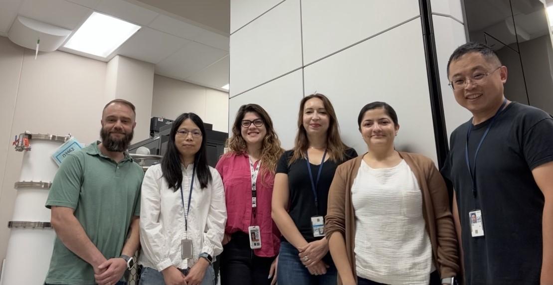
Team
Staff
Principal Investigators overseeing the facility operation

Forms
This site contains Microscope Access Workflow, proposal templates for access to Krios G4 SPA and Talos Arctica, and Reviewer Evaluation Form. Users can download the forms from the following links:
Publications
Selected Publications (2023 - June 2025)
2025
Modularity of Zorya defence systems during phage inhibition, Nature Comms.16:2344. 2025. Mariano, G., Deme, J.C., Readshaw, J., Grobbelaar, M.J., Frey, A., Bamford, L., Songra, S., Trost, M., Blower, T.R., Palmer, R. and Lea, S.M.
Probabilistic single-particle cryo-EM ab intio 2D reconstruction in SIMPLE, Acta Cryst. D, doi: 10.1107/S2059798325005686, 2025. Van, C.T.S., Reboul, C.F., Caesar, J.E., Meana-Paedan, R., Lountos, G.T., Deme, J.C., Bryant, O.J., Johnson, S., Piczak, C.T., Valkov, E., Lea, S.M. and Elmlund, H.
Structural insights into human topoisomerase 3β DNA and RNA catalysis and nucleic acid gate dynamics. Nature Comms., 16: 834, 2025. Yang, X., Chen, X., Yang, W and Pommier, Y.
Intracellular pathogen effector reprograms host gene expression by inhibiting mRNA decay, Nature Comms., 16: 6452, 2025. Levdansky, Y., Deme, J.C., Turner, D.J., Piczak, C.T., Pekovic, F., Tarasov, S.G., Lea, S.M. and Valkov, E.
HIV-1 vif mediates ubiquitination of the proximal protomer in the APOBEC3H dimer to induce degradation. Nature Comms. 16: 5879, 2025. Skorupka, KA, Matsuoka, K., Hassan, B., Ghirlando, R., Balachandran, V., Chen, T., Walters, KJ, Schiffer, CA, Wolf, M., Iwatani, Y. and Matsuo. H.
2024
Structural basis for antibiotic transport and inhibition in PepT2, Nature Comms. 15:8755, 2024. Parker, J.L., Deme, J.C., Lichtinger, S.M., Kuteyi, G., Biggin, P.C.*, Lea, S.M. and Newstead, S.
The type 9 secretion system caught in the act of transport. Nature Microbiol. 9:1089-1102, 2024. Lauber, F., Deme, J.C., Liu, X., Kjaer, A., Miller, H.L., Alcock, F., Lea, S.M. and Berks, B.C.
Structural basis of directional switching by the bacterial flagellum. Nature Microbiol. 9:1282-1292. Johnson, S., Deme, J.C., Furlong, E.J. and Lea, S.M.
Structural insights into the cooperative nucleosome recognition and chromatin opening by FOXA1 and GATA4, Mol. Cell, 84(16):3061 – 3079, 2024. Zhou, BR, Feng, H, Huang, F, Zhu, I, Portillo-Ledesma, S, Shi, D, Zaret, KS, Schlick, T, Landsman, D, Wang, Q, Bai, Y
2023
A type VII-secreted lipase toxin with reverse domain arrangement. Nature Comms. 14:8438 PMC10730906. 2023. Garrett, S.RR., Mietracjh, N., Deme, J.C., Bitzer, A., Yang, Y., Ulhuq, F.R., Kretschmer, D., Heilbronner, S., Smith, T.K., Lea, S.M. and Palmer, T.
Structure and assembly of the NOT10:11 module of the CCR4-NOT complex. Comms Biol. 739: doi: 10.1038/s42003-023-05122-4.PMC10352241. 2023. Levdansky, Y., Raisch, T., Deme, J., Pekovic, F., Elmlund, H., Lea, S.M. and Valkov, E.
Review Committee
Ping Zhang (NCI)
Hans Elmlund (NCI)
Jinwei Zhang (NIDDK)
Di Xia (NCI)
Doreen Matthies (NICHD)
Jiansen Jiang (NHLBI)
Rick Huang (NCI)
Yawen Bai (NCI)
Ulrich Baxa (NIDDK)
Naoko Mizuno (NHLBI)
Eugene Valkov (NCI)
