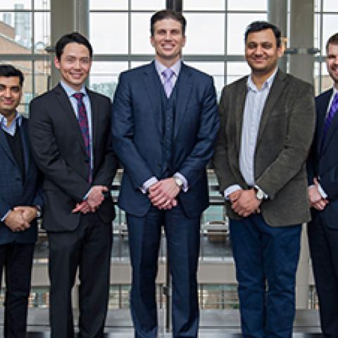
CCR researchers and clinicians with world-class expertise in a wide range of disciplines have started a clinical program aimed at understanding how liver metastases form in colorectal cancer, which may shed light on how to treat or even prevent them. Pictured here are Dr. Hernandez and his team.
Photo credit: Marleen Van den Neste
Metastasis, or the spread of cancer cells beyond the site of the initial tumor, is the leading cause of death in cancer patients. Despite this fact, the mechanisms underlying metastasis formation are still not entirely understood. To better understand metastatic disease – and how to treat or even prevent it – Jonathan Hernandez, M.D., Investigator in the Thoracic and Gastrointestinal Oncology Branch, and colleagues have launched a clinical program comprised of multiple studies investigating metastatic colorectal cancer. The studies center around the group’s ability to keep tumor-containing liver alive after it is removed from a patient.
“We will be able, for the first time, to keep tumor-bearing liver viable outside of the body for novel drug testing and tracking of patients’ individual tumor cells,” says Hernandez. “This is the first time anything like this has been done.”
Demonstrating CCR’s unparalleled opportunities for multidisciplinary collaboration, the new clinical program brings together 10 researchers and clinicians with expertise in medical/surgical oncology, pharmacology, hepatology, pathology, advanced imaging and interventional radiology. Thanks to the unique patient-centered, research-oriented environment of CCR, found at few if any other institutions, “something like this would likely not happen anywhere else in the world,” Dr. Hernandez says.
In many cases of colorectal cancer, the disease spreads to the liver. Standard treatment involves chemotherapy and possible surgery, but in many cases, surgery can’t completely remove all of the tumors. A device called a hepatic artery infusion pump (HAIP), which delivers high doses of chemotherapy directly to the liver, has shown promise in colorectal cancer patients with inoperable liver metastases by reducing their growth and shrinking them in many instances so they can be more easily removed during an operation. As part of CCR’s new clinical program, Dr. Hernandez will lead a phase II trial of HAIP therapy for such patients with recruitment starting soon.
The study will test a personalized medicine approach that will involve analyzing the tumors’ genetic profiles to generate a list of novel therapies that are most likely to be effective. Participants who respond to the HAIP therapy will undergo a second surgery to remove their metastases. During this procedure, the researchers will remove all of the remaining tumors with part of the surrounding liver and keep the tumor-containing liver alive in the laboratory instead of immediately sending it to pathologists. They will then test the therapies predicted to work—based on the tumors’ genetic profiles—on these live tissue samples. Should participants’ tumors recur, the results from these experiments will be used to guide additional therapy.
“Live tumor-containing tissue is an ideal model system,” Dr. Hernandez says. Unlike a patient-derived xenograft, a common tumor model that involves growing a tumor transplanted from a patient inside a mouse, this is the actual tumor in its native environment with its native stroma, all of which plays a crucial role in disease.
If successful, this approach may streamline drug development. Instead of investing time and resources in clinical trials that test the same treatment in every patient, researchers could personalize treatment and test it in models of patients’ own tumors before administering it to patients themselves. “This has the potential to propel personalized medicine forward decades,” Dr. Hernandez says.
The team also seeks to take a preventative approach to metastases. Dr. Hernandez and his team believe that blocking growth of the individual tumor cells that have escaped to distant organs like the liver may be a very effective strategy for patients undergoing surgery to remove all tumors. When tumor cells land on a distant organ, they often lie dormant before they begin to grow and form metastases—but current methods to image tumors, such as MRI and CT scans, can’t detect individual tumor cells.
“Metaphorically speaking, we are trying to keep the hibernating grizzly bear asleep rather than trying to manage it once it’s awake and hungry. Until now, we simply couldn’t find the grizzly bears in order to figure out how to keep them hibernating”.
His team will break down a given patient’s tumor into individual cells after it is removed, label those individual cells with a tracking device and inject the cells into that patient’s liver tissue being kept alive outside the patient’s body. Using fluorescent-label multiphoton microscopy, "the cells can be tracked in real-time so that we can begin, for the first time, to understand this process and think about what therapies can prevent these cells from growing.”
Dr. Hernandez says that even metastatic colorectal cancer patients who aren’t enrolled in the HAIP clinical trial could donate their tissue and participate in the clinical program. Hernandez also envisions the scope of the program expanding to look at metastases in other organs and types of cancer to hopefully find new options for patients fighting metastatic disease.