Joseph M. Ziegelbauer, Ph.D.
- Center for Cancer Research
- National Cancer Institute
- Building 10, Room 6N110
- Bethesda, MD 20892-1868
- 240-858-3267
- ziegelbauerjm@nih.gov
RESEARCH SUMMARY
Our previous research discovered functions of viral microRNAs related to inhibiting apoptosis, evading immune responses, and promoting angiogenesis. Ongoing research focuses on networks and pathways redundantly targeted by multiple viral microRNAs and circular RNAs. In collaboration with Drs. Yarchoan, Ramaswami, Lurain, and Krug in HAMB, we have been investigating human and viral gene expression patterns in normal tissue, in Kaposi sarcoma (KS), and other KSHV-infected pathological tissues. We are using a variety of expression profiling techniques and functional studies to determine the importance of these gene expression patterns. These studies will lead to the discovery of new therapeutic targets for inhibiting viral oncogenesis.
Areas of Expertise

Joseph M. Ziegelbauer, Ph.D.
Research
Kaposi's sarcoma-associated herpesvirus (KSHV) is the responsible agent for Kaposi's sarcoma (KS), primary effusion lymphoma and multicentric Castleman's disease. KSHV expresses multiple microRNAs that modulate human gene expression. Previously, we have discovered and published around 40 validated cellular mRNA targets of Kaposi's Sarcoma Herpesvirus (KSHV) miRNAs. Recently, we have started studying circular RNAs, which are formed by back-splicing events, lack poly-A tails, can regulate gene expression, and are more stable than linear mRNAs. Circular RNAs have recently been shown to inhibit specific miRNAs and have other activities. Our goals have been: 1. To identify circular RNAs that change in expression with viral infection. 2. To discover new viral circular RNAs and understand their biogenesis. 3. To determine the functions and mechanisms of human and viral circular RNAs. We previously reported that Kaposi's sarcoma herpesvirus (KSHV) infection alters the expression of hundreds of human circular RNAs. We showed that a human circRNA, hsa_circ_0001400 (circ_0001400, circRELL1), is induced by various pathogenic viruses, namely KSHV, Epstein-Barr virus, and human cytomegalovirus. Our findings that circ_0001400 levels increase upon infection with multiple human herpesviruses suggests a common mechanism shared across diverse viral infections. However, how these viruses regulate circRNA transcript levels remains elusive. We observed that while circ_0001400 increases during infection, the mRNA from the same gene locus, RELL1, does not show similar regulation. This discrepancy led us to postulate that infection changes of circRNAs occur co-/post-transcriptionally.
We also characterized the circRNAome of HSV-1, KSHV, and MHV68 infections. We used cell culture and mouse models (with our collaborators) to thoroughly profile circRNAs expressed during lytic or latent phases. To facilitate high-throughput analysis of circular transcripts, we developed a custom bioinformatic pipeline called circRNA DAQ (Detection, Annotation, Quantitation). To confirm that predicted transcripts were truly circular, we complemented sequencing approaches with RNase R digestion assays and divergent primer amplification. Through these approaches, we identified thousands of viral circRNAs, some of which approach the abundance of the housekeeping gene GAPDH. It is exciting to note that we are the first to identify HSV-1 encoded circRNAs, including circular transcripts derived from the latency-associated transcript (LAT). Next, we characterized cis- and trans-acting factors that promote circRNA biogenesis. We found that viral circRNA synthesis was resistant to major spliceosome inhibition and not reliant on canonical splice donor-acceptor sites. During lytic infection, viral circRNAs tiled the entire genome, with the late phase of lytic transcription promoting rampant back-splicing. Additionally, using eCLIP and Nascent (4SU) RNA-Seq, we determined that the KSHV RNA binding protein (ORF57) enhanced viral circRNA accumulation post-transcriptionally.
Our work has identified thousands of novel herpesvirus transcripts and elucidated a unique splicing mechanism driven by lytic replication. As viral circRNAs were completely unstudied until the last six years, our work lays the foundation for a novel class of molecules that may contribute to herpesvirus replication, persistence, and tumorigenesis. Importantly, since circRNAs appear to be ubiquitously expressed across assorted virus families, our findings have implications for other viral pathogens and associated diseases.
In collaboration with Drs. Yarchoan, Ramaswami, Lurain, and Krug in HAMB, we have been investigating human and viral gene expression patterns in normal tissue, in Kaposi sarcoma (KS), and other KSHV-infected pathological tissues. Little is known about cellular and viral RNA transcripts in KS lesions and how these factors are influenced by prior therapy, concurrent KSHV-associated diseases, or the tissue location of KS lesions. We sought to understand how human gene expression and viral transcripts are altered in KS as compared to normal tissue using matched patient samples. In this study of participants with well-annotated clinical characteristics, we also investigated both skin and GI KS to determine differences in the viral and host transcripts between these lesions. In our study, we analyzed samples from 19 participants, including both cutaneous and gastrointestinal (GI) Kaposi's sarcoma (KS) samples. The majority of these individuals were coinfected with HIV, and some presented other conditions such as KICS, MCD, and PEL. Upon analysis, we found that GI KS samples had a higher viral load (VL) for KSHV than the skin KS samples, and similarly, HIV VL was also higher in individuals with GI KS. The CD4+ T cell counts were however, comparable in both types of KS samples.
In investigating gene expression patterns, we discovered significant differences in differentially expressed genes (DEGs) between the two types of KS lesions. We identified 370 DEGs unique to skin KS, 58 DEGs unique to GI KS, and 26 common to both compared to their normal tissues. Notably, we found enrichment in IL-6 signaling, HIFα signaling, and the granulocyte adhesion and diapedesis pathway in both locations of KS. The B cell receptor signaling pathway, however, was only enriched in the skin KS samples. There were also some genes that showed distinct patterns in GI and skin KS, such as IL1A, which was upregulated in GI KS but repressed in skin KS. Overall, our study revealed significant differences in gene expression in skin and GI KS lesions, providing valuable insights into the pathogenesis of Kaposi Sarcoma. The differential gene expression in these lesions may help guide future research and treatment approaches.
Publications
- Bibliography Link
- View Dr. Ziegelbauer's PubMed Summary.
Noncanonical circRNA biogenesis driven by alpha and gamma herpesviruses
A virus-induced circular RNA maintains latent infection of Kaposi’s sarcoma herpesvirus
Transcriptional landscape of Kaposi sarcoma tumors identifies unique immunologic signatures and key determinants of angiogenesis
Discovery of Kaposi's sarcoma herpesvirus-encoded circular RNAs and a human antiviral circular RNA
Viral MicroRNAs Repress the Cholesterol Pathway, and 25-Hydroxycholesterol Inhibits Infection
Biography

Joseph M. Ziegelbauer, Ph.D.
Dr. Joseph Ziegelbauer received a B.S. from Cornell University with a major in biology and focus on biochemistry while working in the laboratory of Dr. John Lis. He then joined the laboratory of Dr. Robert Tjian for his graduate research where he received his Ph.D. from the University of California - Berkeley. Later, he began studying viral microRNAs in the laboratory of Dr. Don Ganem at the University of California - San Francisco. Dr. Ziegelbauer joined NCI in November 2008 and received tenure in 2019. He was appointed Deputy Branch Chief in 2023. His previous focus was elucidating the functions of microRNAs expressed by Kaposi's sarcoma-associated herpesvirus (KSHV or HHV-8). Currently, he is studying circular RNAs and using single-cell and spatial transcriptomic techniques to understand viral pathogenesis in patient samples.
Job Vacancies
We have no open positions in our group at this time, please check back later.
To see all available positions at CCR, take a look at our Careers page. You can also subscribe to receive CCR's latest job and training opportunities in your inbox.
Team
News
Learn more about CCR research advances, new discoveries and more
on our news section.
Lab Life
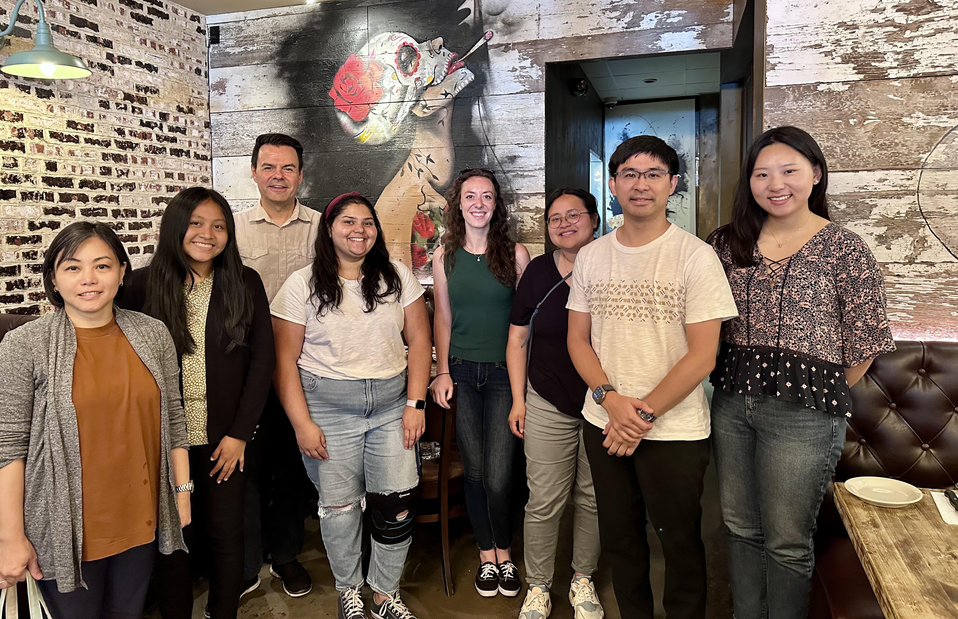
Our lab photo from July 2023.
This was in 2025.
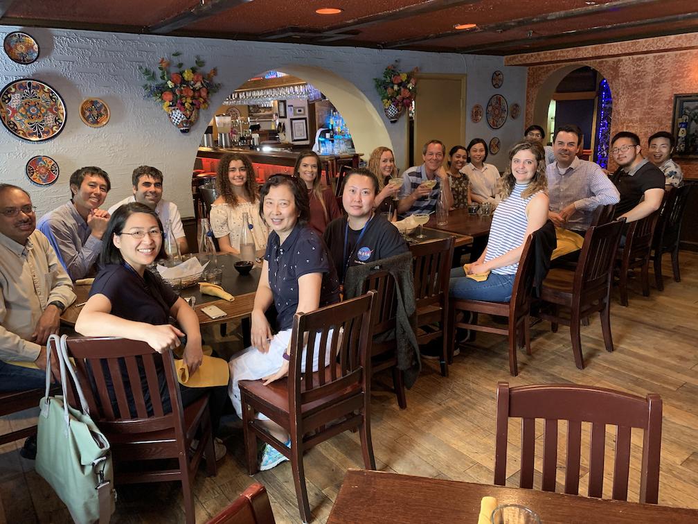
Celebrating Joe's tenure appointment!
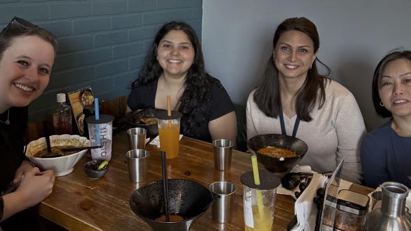
HAMB goes out for lunch.

