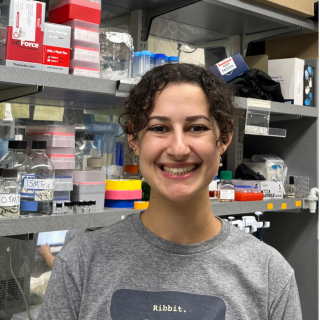
Jaeho Yoon, Ph.D.
- Center for Cancer Research
- National Cancer Institute
- Building 560, Room 22-12
- Frederick, MD 21702-1201
- 301-846-7086
- jaeho.yoon@nih.gov
RESEARCH SUMMARY
Dr. Yoon has been interested in the planar cell polarity (PCP) in his preferred animal model, the developing Xenopus embryo. Polarized cellular behaviors that are governed by PCP signaling represent a vital process for orchestrating cells to form a distinct polarity axis in a tissue. This PCP signaling is driven by various stimuli such as spatial/temporal gene expression, Wnt morphogen gradients and asymmetric subcellular enrichment of PCP proteins. Since abnormal regulation of PCP signaling causes severe birth defects and defects of tissue function, the PCP process has been known as an essential feature for proper embryo development and diverse functions in many tissues. Aberrant Wnt/PCP signaling has been strongly implicated in cancer progression.
Areas of Expertise
Biography

Jaeho Yoon, Ph.D.
Dr. Jaeho Yoon obtained his Ph.D. in developmental biology in the laboratory of Prof. Dr. Jaebong Kim at Hallym University in South Korea in 2012. Dr. Yoon studied the mechanisms of convergence and extension (CE) cell movements in Xenopus gastrula. For his postdoctoral studies, he joined the Cancer and Developmental Biology Laboratory at the National Cancer Institute at Frederick in Dr. Ira Daar’s lab in Dec 2013 as a Visiting Fellow. His research primarily concentrated on Eph-ephrinB signaling during embryogenesis and its implications in cancer metastasis. He has served as a Staff Scientist in the laboratory since August 2019.
Research
1. Exploring the mechanisms that regulate Planar Cell Polarity.
Numerous studies have delved into the influence of non-canonical Wnt ligands, specifically Wnt 4, 5, and Wnt 11, on planar cell polarity (PCP) during diverse developmental processes. These ligands have been implicated in coordinating PCP elements in various events like the closure of the neural tube, the development of fly wings, the formation of cochlear hair cells, and limb development. However, the contradictory findings, particularly the suggestion that Wnt ligands may not be necessary for establishing PCP in Drosophila wing development, cast doubt on the signals responsible for initiating PCP. Earlier research in amphibian vertebrate embryos has proposed that secreted signaling molecules from the dorsal blastopore lip play a crucial role in instructing PCP. The acquisition of PCP in the neural plate advances from posterior to anterior positions. Recent our studies have revealed a link between the spatial and temporal expression of Wnt4 and this progressive PCP acquisition, indicating Wnt4's potential role as an instructive signal. Furthermore, the loss of Wnt4 results in convergent and extension (CE) cell movement abnormalities and abnormal Vangl2 localization, suggesting Wnt4's critical role in guiding planar cell polarity. This study will define whether Wnt4 is involved in instructing PCP and how cells transport Wnt4 to regulates PCP.
2. Enhancing Tools for Exploring Complex Signaling Pathways in Developing Xenopus Embryos.
In recent years, developmental biology research has made remarkable progress, driven by innovative technologies that enable precise manipulation and visualization of intricate cellular processes. Among these state-of-the-art technologies are proximity labeling and optogenetic tools, which have significantly advanced our understanding of developmental processes. Proximity labeling is a versatile methodology that allows us to map protein-protein interactions and investigate protein localization in live cells with exceptional spatial and temporal resolution. This approach typically involves fusing enzymes like peroxidases or biotin ligases to a protein of interest. These enzymes biotinylate neighboring proteins, marking them for subsequent isolation and analysis. Proximity labeling has found widespread utility across diverse fields, including proteomics, structural biology, neuroscience, and developmental biology. Optogenetics, on the other hand, controls the capabilities of light-sensitive proteins such as opsins and cryptochromes to exert precise control and manipulation over cellular processes with high temporal precision. By genetically introducing these light-sensitive proteins into target cells, we can use light pulses of specific wavelengths to activate or inhibit their functions. Optogenetics has significantly advanced our understanding of neuroscience, cell signaling, and developmental biology. However, these cutting-edge techniques require modification and optimization for Xenopus embryo research. Therefore, our primary goal is to optimize proximity labeling and optogenetic tools to enhance their efficiency in Xenopus embryos. These techniques have the potential for broad applications across various scientific domains and hold promise for further advancements in both fundamental research and clinical applications.
3. EphrinB2/Ror2 Interaction Regulates Neural Tube Closure
During primary neurulation, the neural plate differentiates from the ectoderm layer and the cells begin to migrate, which results in the dorsal bending of the neural plate and the joining of the lateral edges to form the neural tube. The regulation of cell shape and movement during this process is orchestrated by several distinct pathways of which the Eph/ephrin and the Wnt-planar cell polarity (Wnt-PCP) signaling pathways play a critical role. Numerous studies have demonstrated that the knockdown of any one of the Wnt-PCP components causes abnormal cell polarity and migration during neural tube closure. In our previous study, we showed that the expression level of ephrinB2, a component of the Eph/ephrin signaling pathways, is important for proper neural tube and F-actin formation, which is critical for apical constriction and neural tube closure. Nonetheless, little is known about the molecular mechanisms pertaining to ephrinB2 and its involvement in regulating apical constriction during neural tube closure. In this study, we performed an immunoprecipitation-mass spectrometric analysis (IP-MS) using ephrinB2-HA overexpressed in embryos and found Wnt4, a non-canonical Wnt pathway protein, and Ror2, a co-receptor for Wnt-PCP signaling, to be novel binding partners of ephrinB2 during neural tube closure. After performing a protein interaction domain mapping (IDM) assay, Wnt4 was found to interact with the intracellular domain of ephrinB2 while the proline-rich domain (PRD) of Ror2 was found to interact with the PDZ binding motif of ephrinB2. Loss-of-function analyses using ephrinB2 and Ror2 morpholino oligos demonstrated significantly decreased contractile actin bundle formation and caused neural tube closure defects. These defects were subsequently rescued by the re-expression of the wild-type counterparts of ephrinB2 or Ror2 and not by the re-expression of the interaction mutants (ephrinB2-delta PDZ or Ror2-delta PRD). In addition, we found that ephrinB2 interacts with Shroom3, a key regulator for contractile actin bundle formation during neural tube closure. By determining that activation of Wnt4 leads to the downstream interaction between EphrinB2 and Ror2, we shed light on the molecular mechanisms of genes that are involved in neural tube morphogenesis. Our study can advance our understanding of neural tube defects (NTDs), which are the second most common birth defect in humans. Moreover, we have gained mechanistic insight into a pathway that plays a substantive role in cancer progression.
4. Dual Transcriptomics – Proteomics of Developing Cranial Neural Crest Cells
A detailed understanding of the complex genetic networks that regulate cranial neural crest development is important because of its implications for multiple cell lineages and human diseases, including cancer metastasis. Cranial neural crest (CNC) cells are a migratory stem cell-like population having a limited capacity for self-renewal. It has been clearly established that CNCs use guidance cues and the local extracellular matrix to collectively migrate. However, a thorough mechanistic understanding of CNC collective migration and how it varies between subpopulations of CNCs progressing to different locations and fates is still lacking. A significant number of studies on metastatic cancer cells reveals remarkable parallels to CNC epithelial-to-mesenchymal-transition (EMT) and migration. Our work supports the parallels between metastasis and CNC migration. EphB/ephrinB proteins are highly expressed in specific CNC arches, and a number of studies have shown that EphB/ephrinB interactions are metastasis suppressive. How cancer cells influence the micro-environments and conditions that make EMT and migration possible is an area of intense interest and will likely provide mechanistic insight into both the mechanisms of tumorigenesis and the plasticity of CNCs. The migrating CNC cells form four branchial arches during development. Each branchial arch develops into specific bones, muscles and nerve cells, which are highly conserved in vertebrates. The disparate gene expression patterns between these groups of cells remain poorly understood. The different transcriptomes of each of the branchial arches have not been analyzed in detail, and specific gene expression signatures in the branchial arches remain unclear. In order to identify specific genetic signatures of each of the branchial arches, we will analyze the gene expression profiles of each branchial arch using both single cell RNA-seq and single cell mass spectrometry. This study will define the expression and signaling response features of distinct CNC streams, and will likely reveal critical players involved in cancer cell migration and invasion.
Publications
- Bibliography Link
- View Dr.Yoon's PubMed Summary.


