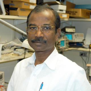
Nallathamby Devasahayam, B.S.
- Center for Cancer Research
- National Cancer Institute
- Building 10, Room B3B69
- Bethesda, MD 20892
- 301-402-6320
- devasahn@mail.nih.gov
RESEARCH SUMMARY
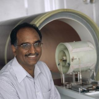
Dr. Devasahayam developed the radiofrequency (300 - 750 MHz) pulsed electron paramagnetic resonance (EPR ) imaging system currently in use in the Biophysical Spectroscopy and Imaging Section. His paper, “Parallel Coil Resonators for Time-Domain Radiofrequency Electron Paramagnetic Resonance Imaging of Biological Objects,” published in the Journal of Magnetic Resonance (142:168-76, 2000) was the first whole-body mouse EPR image and was featured on the cover. Since then, Dr. Devasahayam has completed many software and hardware enhancements to continuously improve mouse imaging. To date, he has 8 patents in Fourier Transform EPR (FT-EPR) imaging techniques. Efforts are now underway to scale up EPR imaging techniques for human applications.
Areas of Expertise

Nallathamby Devasahayam, B.S.
Publications
Parallel coil resonators for time-domain radiofrequency electron paramagnetic resonance imaging of biological objects
Evaluation of a high-speed signal-averager for sensitivity enhancement in radio frequency Fourier transform electron paramagnetic resonance
Tailored sinc pulses for uniform excitation and artifact-free radio frequency time-domain EPR imaging
Strategies for improved temporal and spectral resolution in in vivo oximetric imaging using time-domain EPR
A novel programmable pulse generator with nanosecond resolution for pulsed electron paramagnetic resonance applications
Biography

Nallathamby Devasahayam, B.S.
Nallathamby Devasahayam started his engineering career in 1971 at the Indian Institute of Technology Madras (IIT/M), Chennai, India. He worked there in defense related research projects, microprocessor and computer labs. He also worked in the Regional Sophisticated Instrumentation Center of IIT/M, where he was in charge of development and maintenance of several sophisticated analytical and research spectroscopic instruments. In that center he also developed an X-Band Pulsed Electron Paramagnetic Resonance (EPR) spectrometer. During that period he also visited the laboratory of Prof. Dr. Arthur Schweiger, ETH Zurich to get in depth knowledge in Pulsed EPR techniques. With that, after more than two decades of experience at IIT/M, he joined in 1996 the Bio Physical Spectroscopy and Imaging Section of Radiation Biology Branch, CCR, NCI to develop the Radiofrequency (300 - 750 MHz) Pulsed EPR imaging system currently in use. His paper 'Parallel coil resonators for time-domain radiofrequency electron paramagnetic resonance imaging of biological objects' published in the Journal of Magnetic Resonance, January, 2000 is the first whole body mouse EPR image and was cited at the Cover page. Then onwards many software and hardware developments are being carried out to continuously improve the mouse imaging. To his credit he has eight patents in FT-EPR imaging techniques. Efforts are now under way to scale up the EPR Imaging techniques for human applications.
