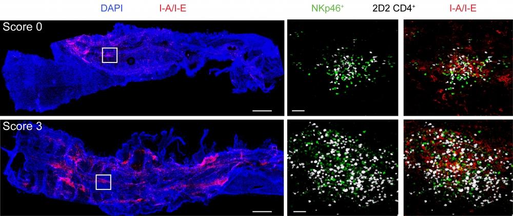
2D2 CD4+ TH17 cells were adoptively transferred into NKp46+ reporter mice (NKp46-Cre+ Rosa26-loxP-STOP-loxP-YFP). Spinal cord meninges were isolated from NKp46+ reporter mice before the onset of symptoms (score 0) and during the peak of disease (score 3) and stained with DAPI and anti-MHCII (I-A/I-E). Inset images show clusters of NKp46+ (green) and 2D2 CD4+ T cells (white), without (left) and with (right) MHCII (I-A/I-E) staining. Scale bars, 500 mm for main images or 40 mm for insets.
