
Philippe Youkharibache, Ph.D.
- Center for Cancer Research
- National Cancer Institute
- Building 10, Room 2B44A
- Bethesda, MD 20892
- 240-858-3181
- philippe.youkharibache@nih.gov

RESEARCH SUMMARY
Dr. Youkharibache’s research aims at: 1) understanding self-association determinants of proteins, especially cell surface and membrane proteins, at several levels of the molecular organization to provide a structural basis for therapeutic protein engineering; 2) developing methods, software, and databases to support the design and engineering of immuno oncology and antiviral therapeutics; and 3) improving the design and engineering of novel immunotherapeutic molecules in clinical trials to treat cancers.
Dr. Youkharibache uses macromolecular structure analysis and computational tools to discover hidden relationships within and between protein domains; he is an expert in macromolecular symmetry analysis. He has inspired, designed and managed the development of numerous successful scientific software packages in both commercial and academic environments, including tools for molecular modeling, computer-aided drug design, structure, and dynamics analysis, next-gen sequencing data analysis, protein structure symmetry analysis, and structural bioinformatics.
His most recent work has been focused on 1) chimeric antigen receptors architecture, and its impact on T-cell function and 2) receptor binding domains from human endogenous retrovirus and zoonotic viruses to provide working hypotheses for the molecular mechanisms underlying biological function of these complex systems.
Areas of Expertise
Philippe Youkharibache, Ph.D.
Research
Dr. Youkharibache’s research is rooted in the study of protein structure and interactions to provide a guide for the design of anti-cancer and antiviral therapeutic proteins. Our lab develops methods and software to analyze proteins and protein assemblies at the structural level and designs chimeric proteins to be used as ligand or receptors to the cell surface and membrane proteins for anti-cancer therapies. We use a host of computational tools to design and simulate these chimeric proteins' behavior alone and in interaction with target protein on the surface of target cells, whether antigens on the cancer cell or viral receptor binding domain (RBD) receptors.
In the last few years, I have focused on the study of self-association determinants of molecular systems, especially proteins, as revealed by their structural symmetries at several levels of molecular organization: molecular assemblies, protein chains, protein domains [1], and protein supersecondary structures (protodomains) [2]. Symmetry and quasi-symmetry reveal a self-organization principle underlying molecular assemblies. Symmetry analysis helps co-relate interacting proteins, interacting domains, and even interacting parts within domains, that we have named "protodomains" [2]. The departure from exact symmetry (quasi- or pseudo-symmetry) reveals a co-evolution mechanism of related structural elements [2,4]. We showed that ca. 20% of protein domains present internal symmetries [3], while over 50% of protein assemblies present quaternary symmetries. These numbers are even higher among membrane proteins [4] and cell surface proteins, especially those formed from immunoglobulin domains [2], at the heart of immune synapses. Many of the cell surface protein receptors, from T cells to their target cells (TCRs, CD4, CD8, CD28, CTLA4, PD1, PDL1, etc.), are composed of Ig domains interacting through oligomeric pseudo-symmetric arrangements that can be used to design more specific agents. Symmetry analysis offers ways to approach the modulation of function either by enforcing or breaking the symmetry. At a practical level, symmetry can be used to organize data by defining a hierarchical order that relates the information to itself (for a given system) and other systems. We are currently exploring data organization principles and uses as a founding criterion applying artificial intelligence (AI) methods to identify and harness molecular properties to optimize biological function
We develop methods and software to study and design immunotherapeutic proteins. The iCn3D software [5] has been used to analyze and capture knowledge on determinants (molecular interactions) of protein folding and protein assembly that we can then use to design chimeric proteins to interact with target receptors on the surface of cells. We actively develop the open-source software iCn3D in collaboration with the National Center for Biotechnology Information (NCBI).
Plasticity is a property of macromolecules, complementary to symmetry. Understanding plasticity determinants in a fold is key to understanding biological function. We are developing an algorithm that can identify key regions of plasticity (hinges) and their sequence determinants from protein structure ensembles. We aim to use this knowledge in the design and tuning of targeted biological properties of chimeric proteins.
One of our main focuses is on chimeric antigen receptors (CAR) used in anti-cancer (CAR T cell) immunotherapies in clinical trials at the NCI. We have shown qualitatively the role of chimeric antigen receptors' plasticity in CAR T cell signaling through model building and simulations. Our modeling tools' application helped us characterize anti-CD19 and anti-BCMA CARs using variable CD8 and CD28 hinge components based on flexibility analysis [6], suggesting that CAR toxicity can be related to its flexibility. Our exploration of CAR extracellular domains by X-ray crystallography techniques resulted in the first report on the formation of a spontaneous rearrangement of a CAR scFv [7] mediated by quasi-symmetry [2], resulting in an unexpected VL-VL arrangement [7]. These early promising results support the development of a pipeline for the rapid modeling of CARs at different scales of complexity and exploring their properties through computational techniques. These computational analyses and new developments are exploring approaches to in-silico structure analysis, mining of big data repositories, new data science methods and machine learning tools [1,2,3,4] to optimize structural and functional properties of chimeric proteins for specific therapies.
More recently, we started applying our methods and software to study binding determinants of RBDs of beta coronaviruses (SARS-CoV-2 and homologs) [8], those of animal and human retroviruses, and of human endogenous retrovirus (HERV) homologs, to their cognate cell surface receptors for the development and optimization of biomarkers and anti-cancer and antiviral therapeutics.
References
[1] Youkharibache, Philippe, Stella Veretnik, Qingliang Li, Kimberly A. Stanek, Cameron Mura, and Philip E. Bourne. 2019. “The Small β-Barrel Domain: A Survey-Based Structural Analysis.” Structure 27 (1): 6–26.
[2] Youkharibache, Philippe. 2019. “Protodomains: Symmetry-Related Supersecondary Structures in Proteins and Self-Complementarity.” Methods in Molecular Biology 1958: 187–219.
[3] Myers-Turnbull, Douglas, Spencer E. Bliven, Peter W. Rose, Zaid K. Aziz, Philippe Youkharibache, Philip E. Bourne, and Andreas Prlić. 2014. “Systematic Detection of Internal Symmetry in Proteins Using CE-Symm.” Journal of Molecular Biology 426 (11): 2255–68.
[4] Youkharibache, Philippe, Alexander Tran, and Ravinder Abrol. 2020. “Pseudo-Symmetric Assembly of Protodomains as a Common Denominator in the Evolution of Polytopic Helical Membrane Proteins.” Journal of Molecular Evolution 88 (4): 319–44.
[5] Wang, Jiyao, Philippe Youkharibache, Dachuan Zhang, Christopher J. Lanczycki, Renata C. Geer, Thomas Madej, Lon Phan, et al. 2020. “iCn3D, a Web-Based 3D Viewer for Sharing 1D/2D/3D Representations of Biomolecular Structures.” Bioinformatics 36 (1): 131–35.
[6] Brudno JN, Lam N, Vanasse D, Shen YW, Rose JJ, Rossi J, Xue A, Bot A, Scholler N, Mikkilineni L, Roschewski M, Dean R, Cachau R, Youkharibache P, Patel R, Hansen B, Stroncek DF, Rosenberg SA, Gress RE, Kochenderfer JN. 2020. “Safety and Feasibility of Anti-CD19 CAR T Cells with Fully Human Binding Domains in Patients with B-Cell Lymphoma.” Nature Medicine 26 (2): 270-280.
[7] PDBid: 7JO8. Cheung, J., Hendrickson, W.A., Kochenderfer, J.N., Youkharibache, P. Crystal structure of a Chimeric Antigen Receptor (CAR) scFv domain rearrangement forming a VL-VL dimer. 2020.
[8] Youkharibache, Philippe, Raul Cachau, Tom Madej, and Jiyao Wang. 2020. “Using iCn3D and the World Wide Web for Structure-Based Collaborative Research: Analyzing Molecular Interactions at the Root of COVID-19.” bioRxiv : The Preprint Server for Biology, July.
Publications
The Small β-Barrel Domain: A Survey-Based Structural Analysis
Protodomains: Symmetry-Related Supersecondary Structures in Proteins and Self-Complementarity
Systematic detection of internal symmetry in proteins using CE-Symm
Pseudo-Symmetric Assembly of Protodomains as a Common Denominator in the Evolution of Polytopic Helical Membrane Proteins
iCn3D, a web-based 3D viewer for sharing 1D/2D/3D representations of biomolecular structures
Biography

Philippe Youkharibache, Ph.D.
Dr. Philippe Youkharibache has a broad background in macromolecular structure analysis, drug design, and data science (informatics), having worked in industry, in academia, and in government research institutions. He did his Ph.D. (Doctorat es-science physiques) at the University Paris-Sorbonne in theoretical and computational chemistry while working at the Ecole Polytechnique in Prof. Fetizon’s Laboratory. He then went to Columbia University for a postdoc in computational biology and molecular modeling with Profs. Cyrus Levinthal and Barry Honig. He played key roles during the rapid rise of the molecular modeling and computer-aided drug design (CADD) software industry. He was a member of several startups, including Polygen started by Prof. Martin Karplus in Waltham. MA and Paris, France, and then Biosym, started by Prof. Arnold Hagler in San Diego, where he developed award-winning software. When both companies were acquired by Corning Life Sciences and merged to become Accelrys, and now Biovia, he moved on to start InPharmatics, a multi-disciplinary Life Sciences informatics company in San Diego. He then focused on subjects related to biodiversity and biodiversity informatics, organizing research collaborations between developing countries rich in biodiversity and pharma companies to identify novel natural products. After returning to France, he participated in the birth of yet another innovative industry on Next-Gen Sequencing data analysis software with Genomatix, a startup company based in Munich, Germany and Paris, France, later acquired by Precision Genetics. Dr. Youkharibache pursued many academic scientific collaborations to facilitate the development of his projects in industry, especially with the PDB (RSCB) in San Diego. Dr. Youkharibache had a keen interest in symmetry as an organizing criterion and a macromolecular organization's fundamental property throughout his career. He furthered his interest on the application of symmetry-related concepts to macromolecular studies during a sabbatical at the San Diego Super Computer Center in Dr. Phil Bourne's lab, where he focused on the identification of protein domain symmetries that guided Ph.D. students and postdocs in that lab in developing CE-Symm, the reference software for molecular symmetry detection, leading to a seminal paper surveying protein domain symmetry in the Protein Data Bank (Myers-Turnbull, et al. 2014). In 2014 Dr. Youkharibache joined Dr. Bourne at the NIH who was heading the Data Science effort in the Office of the Director, to follow up his research at NCBI and start bridging the gap between gene sequence and protein structure with the goal of identifying unique and actionable fingerprints to aid emerging efforts towards precision medicine. He then joined the newly formed Cancer Data Science Lab at the NCI, headed by Dr. Eytan Ruppin, where he leads a team of collaborators across institutes in developing methods, algorithms, software and computational servers aimed at the rational design of immunotherapeutic proteins in collaboration with CCR principal investigators.
Job Vacancies
We have no open positions in our group at this time, please check back later.
To see all available positions at CCR, take a look at our Careers page. You can also subscribe to receive CCR's latest job and training opportunities in your inbox.
Team

Covers
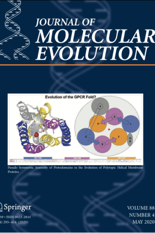
Pseudo-Symmetric Assembly of Protodomains as a Common Denominator in the Evolution of Polytopic Helical Membrane Proteins
Abstract
The polytopic helical membrane proteome is dominated by proteins containing seven transmembrane helices (7TMHs). They cannot be grouped under a monolithic fold or superfold. However, a parallel structural analysis of folds around that magic number of seven in distinct protein superfamilies (SWEET, PnuC, TRIC,FocA, Aquaporin, GPCRs) reveals a common homology, not in their structural fold, but in their systematic pseudo-symmetric construction during their evolution. Our analysis leads to guiding principles of intragenic duplication and pseudo-symmetric assembly of ancestral transmembrane helical protodomains, consisting of 3 (or 4) helices. A parallel deconstruction and reconstruction of these domains provides a structural and mechanistic framework for their evolutionary paths. It highlights the conformational plasticity inherent to fold formation itself, the role of structural as well as functional constraints in shaping that fold, and the usefulness of protodomains as a tool to probe convergent vs divergent evolution. In the case of FocA vs. Aquaporin, this protodomain analysis sheds new light on their potential divergent evolution at the protodomain level followed by duplication and parallel evolution of the two folds. GPCR domains, whose function does not seem to require symmetry, nevertheless exhibit structural pseudo-symmetry. Their construction follows the same protodomain assembly as any other pseudo-symmetric protein suggesting their potential evolutionary origins. Interestingly, all the 6/7/8TMH pseudo-symmetric folds in this study also assemble as oligomeric forms in the membrane, emphasizing the role of symmetry in evolution, revealing self-assembly and co-evolution not only at the protodomain level but also at the domain level.
Pseudo-Symmetric Assembly of Protodomains as a Common Denominator in the Evolution of Polytopic Helical Membrane Proteins. Youkharibache P, Tran A, Abrol R. J Mol Evol. 2020 May;88(4):319-344. doi: 10.1007/s00239-020-09934-4. Epub 2020 Mar 18. PMID: 32189026; PMCID: PMC7162841.

Protodomains: Symmetry-Related Supersecondary Structures in Proteins and Self-Complementarity
Abstract
We will consider in this chapter supersecondary structures (SSS) as a set of secondary structure elements (SSEs) found in protein domains. Some SSS arrangements/topologies have been consistently observed within known tertiary structural domains. We use them in the context of repeating supersecondary structures that self-assemble in a symmetric arrangement to form a domain. We call them protodomains (or protofolds). Protodomains are some of the most interesting and insightful SSSs. Within a given 3D protein domain/fold, recognizing such sets may give insights into a possible evolutionary process of duplication, fusion, and coevolution of these protodomains, pointing to possible original protogenes. On protein folding itself, pseudosymmetric domains may point to a "directed" assembly of pseudosymmetric protodomains, directed by the only fact that they are tethered together in a protein chain. On function, tertiary functional sites often occur at protodomain interfaces, as they often occur at domain-domain interfaces in quaternary arrangements. First, we will briefly review some lessons learned from a previously published census of pseudosymmetry in protein domains (Myers-Turnbull, D. et al., J Mol Biol. 426:2255-2268, 2014) to introduce protodomains/protofolds. We will observe that the most abundant and diversified folds, or superfolds, in the currently known protein structure universe are indeed pseudosymmetric. Then, we will learn by example and select a few domain representatives of important pseudosymmetric folds and chief among them the immunoglobulin (Ig) fold and go over a pseudosymmetry supersecondary structure (protodomain) analysis in tertiary and quaternary structures. We will point to currently available software tools to help in identifying pseudosymmetry, delineating protodomains, and see how the study of pseudosymmetry and the underlying supersecondary structures can enrich a structural analysis. This should potentially help in protein engineering, especially in the development of biologics and immunoengineering.
Protodomains: Symmetry-Related Supersecondary Structures in Proteins and Self-Complementarity. Youkharibache P. Methods Mol Biol. 2019;1958:187-219. doi:10.1007/978-1-4939-9161-7_10. PMID: 30945220.